Where on the Neuron Are Nerve Impulses Summed
The generation of action potential in nerves

The 1963 Nobel Prize for Physiology or Medicine was awarded to Sir John Eccles, Alan Lloyd Hodgkin and Andrew Fielding Huxley for their work on the ionic mechanisms involved in excitation and inhibition in the peripheral and central portion of the nerve cell membrane.
Their work was determining between the rival chemical and electrical theories of synaptic transmission, a controversy at the time. However, the decisive victory of the chemical theory had to wait for the intracellular recording from neuromuscular junctions and nerve cells by Fatt and Katz.
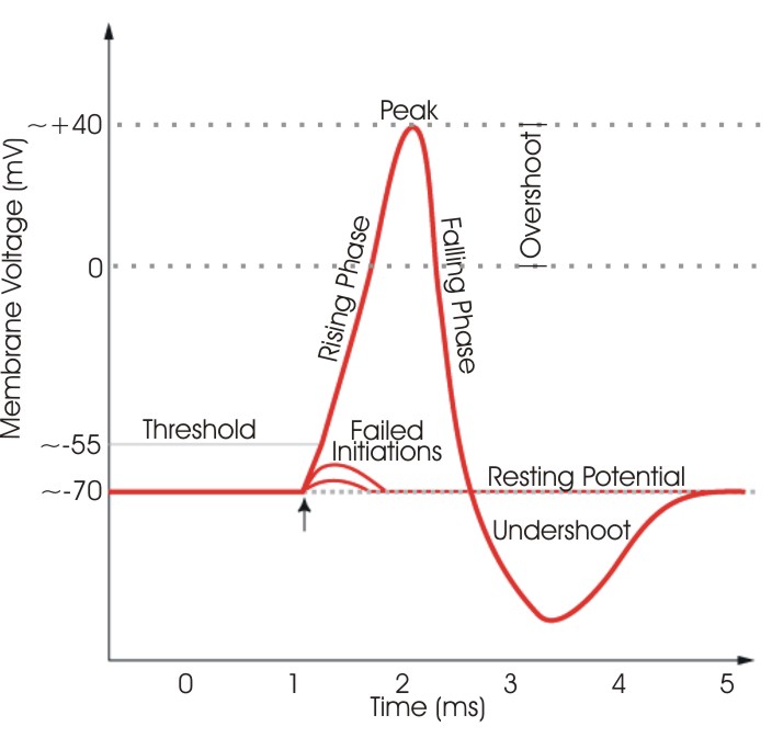
Sir john Eccles studied the fundamental processes of excitation and inhibition of neurons. He used the stretch reflex as a model, easily studied because it consists of two neurons: a sensory neuron synapses in the spinal cord onto a motor neuron. By inserting a capillary microelectrode with a tip diameter of 0.0005 mm into individual nerve cells in the spinal cord, Sir Eccles demonstrated that these cells have a resting potential of about negative 70 millivolts across the cell membrane. If an impulse arriving at the neuron resulted in an excitation, the cell membrane would depolarize (over -70mV), and on the contrary if it caused inhibition, the membrane of the cell would hyperpolarize bellow the resting potential. Single excitatory postsynaptic potentials or EPSPs are not enough to fire an action potential in a nerve. However, the sum of several EPSPs from multiple sensory neurons synapsing onto one motor neuron could cause the motor neuron to fire. On the other hand, inhibitory postsynaptic potentials or IPSPs could subtract from this sum of EPSPs, preventing the motor neuron from firing. Eccles demonstrated in electrical terms the phenomena of neural excitation and inhibition.
His work had fundamental importance in understanding the processing of nerve impulse conduction and helped better understand the nature of ion transfer across nerve cell membranes, further explored by Hodgkin and Huxley.
Both scientists worked together on deciphering the physio-chemical nature of nerve impulses. The nerve impulse is a transmitted electrical event produced by the axon itself when its membrane is depolarized.
During the nerve impulse the action potential exceeds the value of the membrane resting potential by 35% resulting in a membrane potential of about 100 mV that lasts about 1millisecond. The action potential passes like a wave along a nerve and is a fundamental unit in the code by which nerve cells communicate with each other and carry out instruction from the brain for motor and sensory cells. The axons of the nerve cells contain potassium ions kept by membranes from diffusing out into the sodium rich body fluids. The resting potential of the nerve cell is due to a differential electrical potential caused by a concentration build-up of potassium ions within the nerve fibres.
Hodgkin and Huxley observed nerve depolarization in giant squid axons which are about 0.7 mm in diameter, compared to 0.015 mm in vertebrates. They saw that depolarization initiates a leak of sodium ions into the axon, creating a rising phase of nerve impulse. This rise quickly terminates and is replaced by a new leak in potassium ions from the axon during the falling phase. This sodium-potassium balance explains the nerve impulse in quantitative physio-chemical terms.
The contribution to the advancements of knowledge in this field by the three scientists profoundly influenced the research for a wide range of neurological disorders.
- Year: 1963
- Scientist(s): John Eccles, Alan Hodgkin, Andrew Huxley
- Animal(s): Squid
- Countries: Australia, United Kingdom
- Research field(s): Brain and nervous system, Cell biology, Drugs and toxins
- Medical application(s): Basic research
Last edited: 19 November 2014 17:06
Where on the Neuron Are Nerve Impulses Summed
Source: https://www.animalresearch.info/en/medical-advances/nobel-prizes/the-generation-of-action-potential-nerves/